
radiology
Understanding Radiology- How Medical Imaging Helps Diagnose Health Problems
Introduction
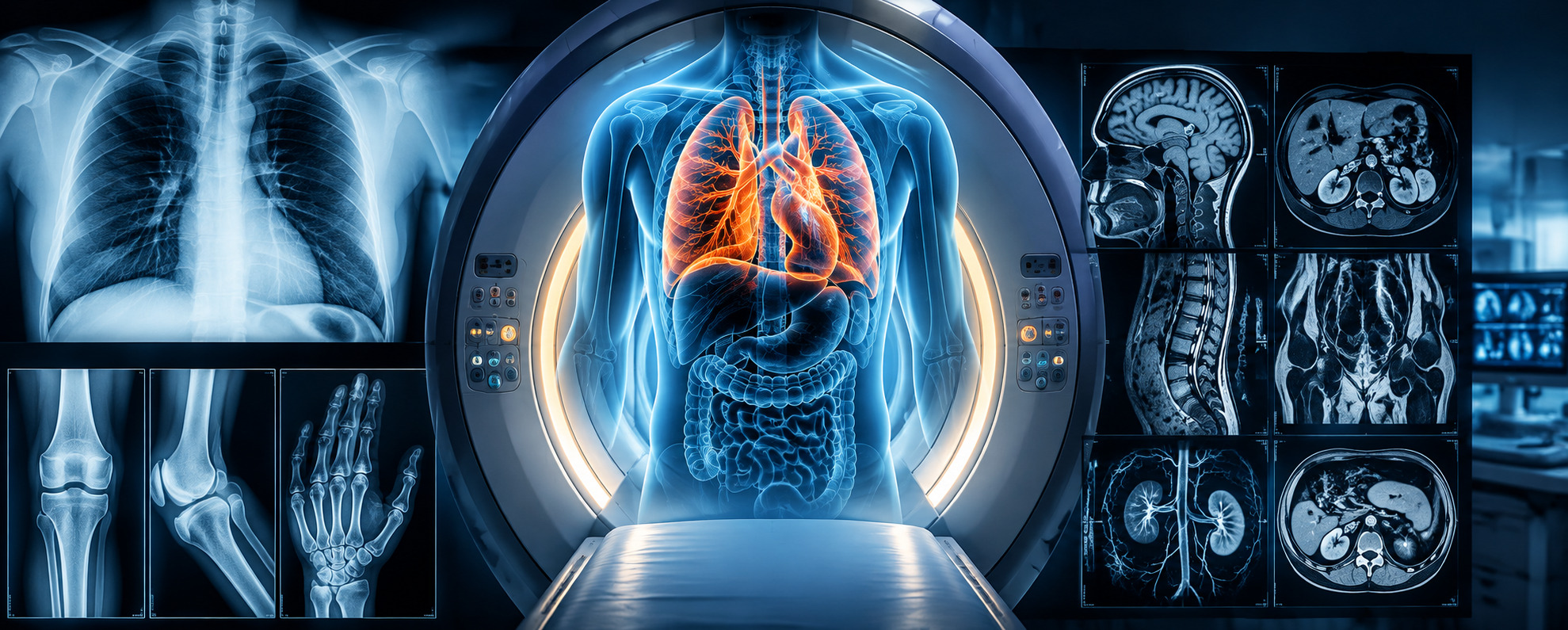
When a doctor needs to understand what is happening inside the body, a physical examination may not always provide enough information. Medical imaging allows doctors to look at bones, organs, blood vessels and other internal structures without surgery.
Radiology is the branch of medicine that uses imaging tests to help doctors diagnose and monitor different health conditions. Depending on the patient's symptoms and the area being examined, the doctor may recommend an X-ray, ultrasound, CT scan or MRI.
The choice of imaging test depends on the patient's symptoms, medical history and the information the doctor needs.
1. What Is Radiology?
Radiology involves using medical imaging to examine different parts of the body. Different imaging tests are used for different purposes.
Common types of medical imaging include:
- X-ray: Commonly used to examine bones and the chest.
- Ultrasound: Uses sound waves to create images of internal organs and tissues.
- CT scan: Uses multiple X-ray images to produce detailed pictures of the body.
- MRI: Uses a magnetic field and radio waves to create detailed images, particularly of soft tissues.
The doctor will choose the most suitable test depending on the patient's symptoms and the condition being investigated.
2. When Might a Doctor Recommend an Imaging Test?
A doctor may recommend an imaging test when more information is needed after discussing the patient's symptoms or carrying out a physical examination.
Medical imaging may be used to:
- Investigate unexplained or persistent pain
- Examine fractures and other bone injuries
- Find the possible cause of abdominal pain
- Investigate certain chest symptoms
- Evaluate headaches and other neurological symptoms
- Examine swelling or changes in internal organs
- Look for signs of infection
- Assess the kidneys and urinary system
- Examine organs and tissues inside the abdomen
Imaging can also be used to monitor a condition that has already been diagnosed and check whether it has changed over time.
Not every symptom requires an imaging test. The doctor will decide whether a scan is appropriate based on the patient's individual condition.
3. What Is the Difference Between X-Ray, Ultrasound, CT and MRI?
Although these tests all produce images of the body, they work differently and provide different information.
X-Ray
X-rays are commonly used to examine bones and the chest. They can help doctors identify fractures and certain abnormalities in the lungs and other areas.
Ultrasound
Ultrasound uses sound waves to produce images of internal structures. It is commonly used to examine abdominal organs, the urinary system and reproductive organs. It is also widely used during pregnancy.
CT Scan
A CT scan uses multiple X-ray images to create detailed cross-sectional images of the body. It can provide more detailed information than a standard X-ray for certain conditions.
MRI
MRI uses a magnetic field and radio waves to produce detailed images. It is particularly useful for examining soft tissues and may be used to assess the brain, spine, joints and various organs.
The doctor will recommend the imaging test that is most appropriate for the health concern being investigated.
4. Is Medical Imaging Safe?
The safety considerations depend on the type of imaging test.
X-rays and CT scans use ionising radiation, while ultrasound and MRI do not use ionising radiation.
Before certain imaging tests, patients may be asked about:
- Pregnancy or possible pregnancy
- Previous reactions to contrast material
- Kidney problems
- Implanted medical devices
- Previous surgeries
- Current medications
Some examinations may also require specific preparation, such as fasting or drinking water before the scan.
It is important to provide accurate information about your health, medications, allergies, pregnancy status and previous medical procedures. This allows the healthcare team to take the necessary precautions before the examination.
5. What Should You Expect During an Imaging Test?
The experience can be different for each type of scan. Some examinations are completed quickly, while others may take longer or require preparation beforehand.
Depending on the examination, you may be asked to:
- Avoid eating or drinking for a specific period
- Remove jewellery or other metal objects
- Change into suitable clothing
- Drink water before certain ultrasound examinations
- Lie or sit in a particular position
- Hold your breath for a short time
- Receive an injection of contrast material, if required
The radiology team will explain the procedure and tell you what you need to do before and during the scan.
If you feel nervous or have questions about the examination, speak with the radiology team before the procedure begins. Following their instructions can help the examination go smoothly.
6. Why Does Choosing the Right Imaging Test Matter?
Medical imaging can provide doctors with important information when diagnosing and monitoring health conditions. However, different scans are designed to show different parts of the body and provide different types of information.
The images are reviewed by a radiologist, who prepares a report based on the findings. The doctor who requested the scan can then use the report along with the patient's symptoms, examination and medical history to decide on the next step.
Depending on the findings, the doctor may recommend:
- Further investigations
- Follow-up imaging
- Medication
- Consultation with another specialist
- Additional treatment
Radiology is therefore an important part of diagnosis and ongoing medical care.
Maruti Hospital provides Radiology services to support the diagnosis and monitoring of various medical conditions.